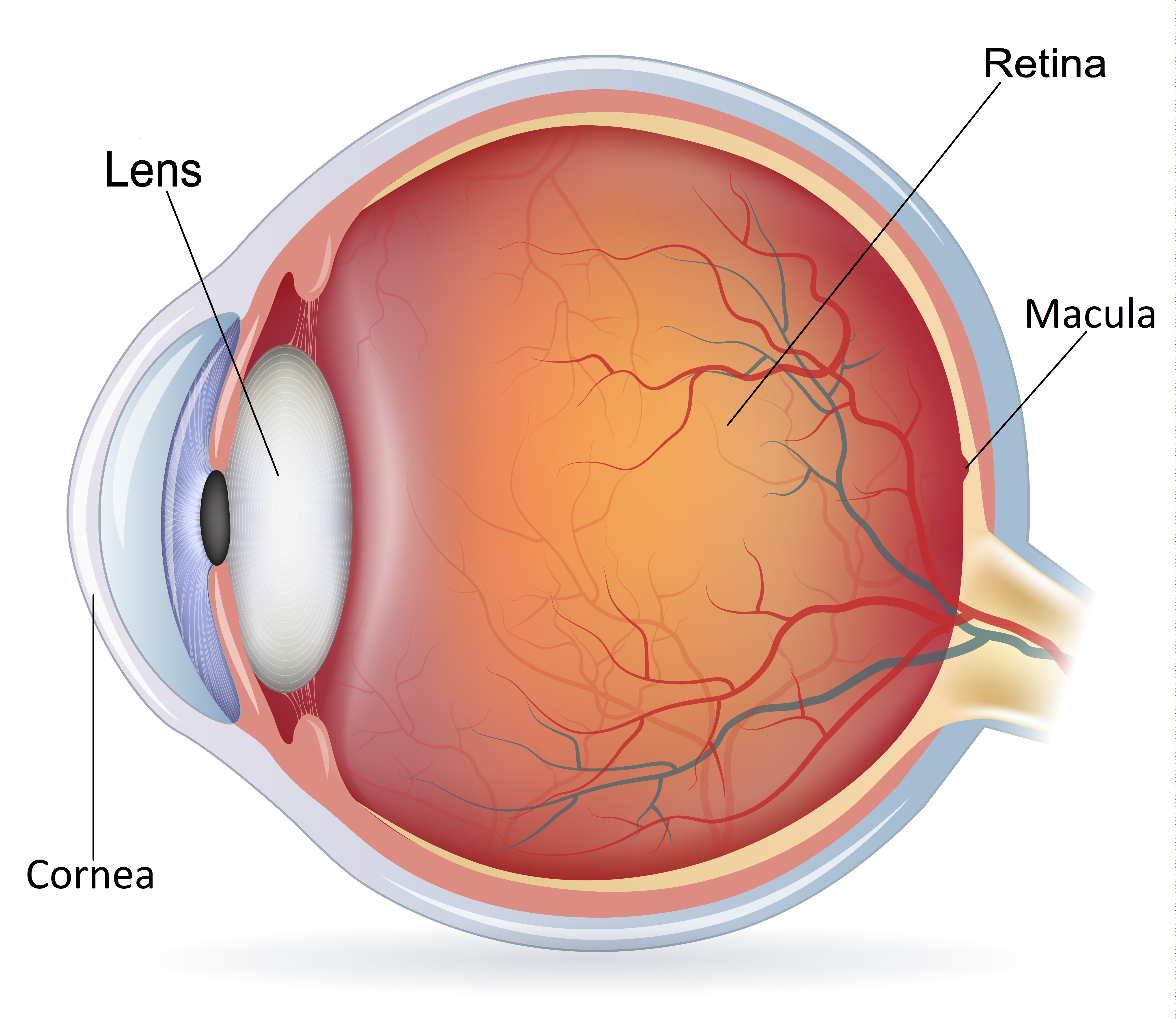
The eye is like a camera with a lens and film in the back of the eye. Between the lens and film, there is a jelly called the vitreous which fills the eye. The retina is the light sensitive film at the back of the eye and a retinal detachment is when the retina peels away from the eye. This is commonly due to a hole or tear forming in the retina causing fluid to pass underneath the retina. Most retinal detachments are often caused by changes that happen to your eye as you age.
The treatment involves surgery with the aim of securing the breaks in the retina and reattaching the retina. The two methods used in surgery are vitrectomy or scleral buckle or a combination of the two. The surgery can be performed under local or general anaesthesia and the decision on the type of anaesthesia is made between you and your surgeon.
A vitrectomy involves removing the vitreous gel (the cause of the tear) from inside the eye. The surgeon then uses either laser or freezing technique to seal the tear. Gas or silicone oil bubble is then inserted into the eye to support the retina while it heals. The gas bubble will slowly self-absorbed typically over 2 to 8 weeks but silicone oil will need an operation to remove it at a later date.

With a gas bubble in the eye, your surgeon may ask you to posture after the operation for up to 7 days. Posturing involves placing your head in a specific position to allow the gas bubble to float into the best position to support the retina, while it reattaches. Posturing after the operation is an important second stage of surgery but if that is not possible and you are worried about coping, please let your surgeon know. You will be required to posture up to 45 - 50 minutes of every hour during the day. The remaining time of every hour can be spent moving around as normal. You must not fly until the gas bubble disappears and you must inform your anaesthetist if you require a general anaesthesia for any operation while there is gas in your eye.
Retinal breaks can also be sealed and supported by stitching a piece of silicone rubber to the outside of the eye. This pushes the eye structures from the outside towards the detached retina, which helps the retina reattach. Laser or freezing technique is also used to seal the tear. The scleral buckle is not visible on the outside of the eye and usually remains in place permanently.
The main aim is to prevent you from going blind in the affected eye. You may have lost vision already due to the retinal detachment and even with successful surgery your vision may not return to normal.
Retinal detachment surgery is not always successful. The success rate for retinal detachment surgery is approximately 80- 90% with a single operation. This means that 2 in 10 people (20%) will need more than one operation. The risks and benefits will be explained to you before you give consent for surgery by your doctor.
Any surgical procedure carries risks which include bleeding, infection however, in retinal detachment surgery this risk is very low (1 in 1000). Although rare, retinal detachment surgery carries the risk of serious consequences, including blindness.
Retinal detachment surgery is a major operation and it is normal to experience some discomfort in the eye following surgery. You will be given specific posturing instructions which need to be followed after surgery. The white of the eye may appear red with swelling to the eyelid. You may have some watering of the eye and a gritty sensation in the eye which slowly disappears in few days. You will be given eye drops to be used for few weeks. You can shower or bath but avoid getting water directly into the eye and abstain from unhygienic environments and anything that puts the eye at risk of injury.
You will be reviewed usually after one week and again after 4 weeks. Vision in the eye will be blurred for a few weeks after the operation but it should improve slowly over time. Your surgeon will discuss your final expected visual outcome of your surgery. If you have any concerns, please discuss this with your surgeon.
You will need rest from work for about two weeks. We advise against driving for the first few weeks. However, any work-related concerns should be discussed with your surgeon.
Understanding retinal detachment surgery can be complicated. This information leaflet may not cover all the concerns you may have about this procedure. Further information can be found at the following websites:
https://www.nhs.uk/conditions/detached-retina-retinal-detachmentThe information mentioned here is based on a variety of sources, including latest published research and the Britain & Eire Association of Vitreoretinal Surgeons.
This must not be used as a substitute for professional medical care by a qualified doctor or other health care professional. Always check with your doctor if you have any concerns about your condition or treatment. We are not responsible or liable, directly or indirectly, for ANY form of damages whatsoever resulting from the use (or misuse) of information contained in this page or found on web pages linked to from this page