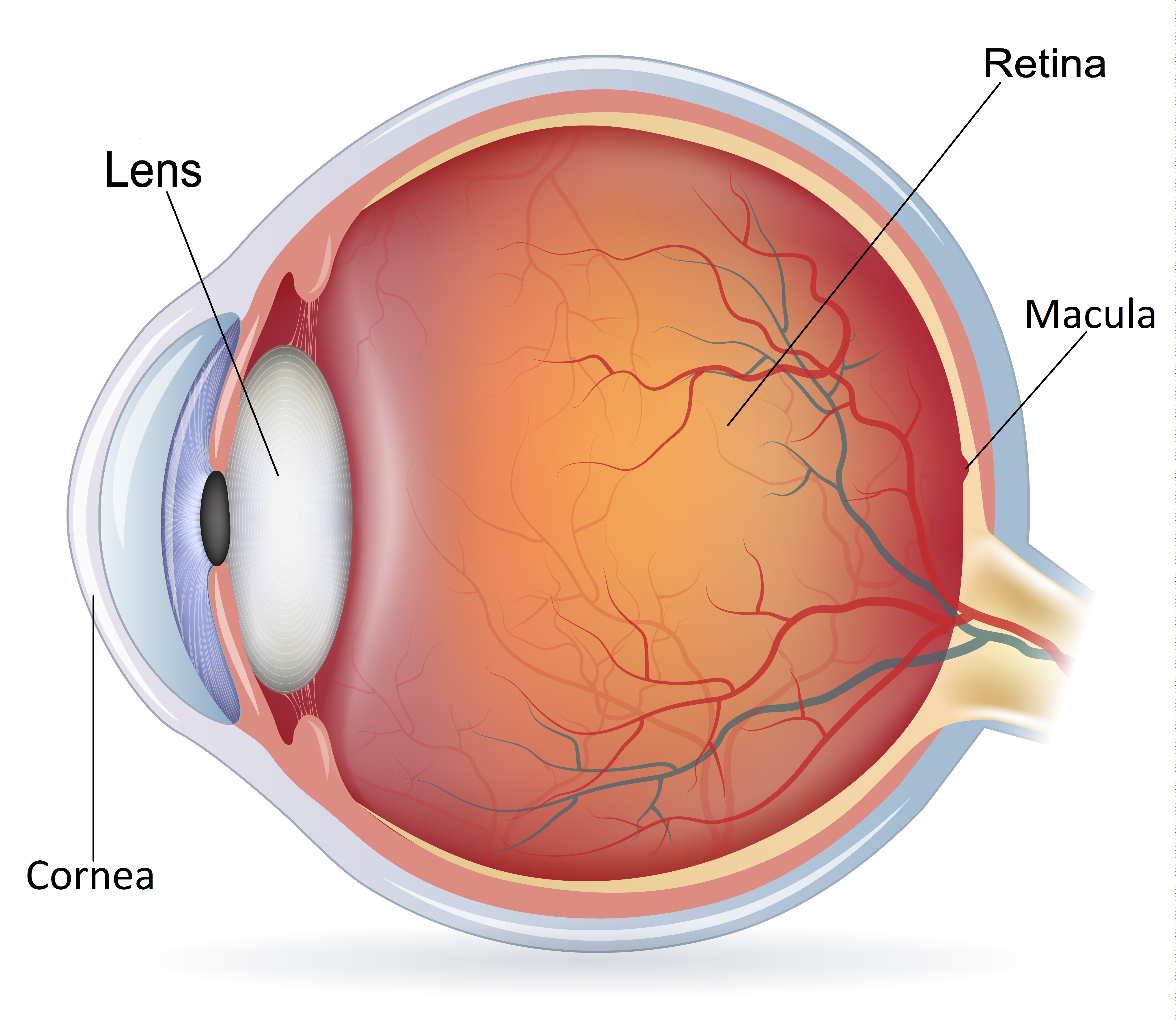
The eye is like a camera with a lens in the front and film in the back of the eye. The retina is the light sensitive film at the back of the eye and the macula is the mostimportant part of the retina, responsible for sharp central vision like reading, driving and focusing objects. A macular hole is the development of a small hole in the centre of the macula.
“Macular holes and macular degeneration are different conditions although they affect the same area of the eye"

We do not know the exact cause of macular holes, but it is believed to be related to the vitreous jelly pulling on the central macula. Macular holes typically occur in patients over 60 years old and tend to affect women more than men. Other factors that can contribute to their development include trauma, being long-sighted or very short-sighted, and having a history of other retinal problems.
The macula is an important area on the retina responsible for sharp central vision. A hole in the macula can cause distorted and blurred central vision, making straight lines appear bent and sometimes resulting in a patch of missing vision in the center. These symptoms become more noticeable when the good eye is covered.
"If left untreated, central vision may deteriorate to the point where even reading large prints, such as the 'Big E' on the eye chart, becomes difficult."


Though a macular hole can be diagnosed by clinical examination, it is necessary to perform an OCT scan of the retina to obtain intricate details of the macular hole and provide advice regarding the best surgical outcome. Mr. Viswanathan has access to a state-of-the-art retinal scanner to investigate macular holes.

The only definitive intervention to treat a macular hole is to do an operation called vitrectomy. Some patients accept the poor central vision and choose not to have surgery. Not having an operation is also an option as there is no right or wrong decision as everyone has different priorities and requirements. You can discuss your decision with Mr Viswanathan regarding whether to have or not to have an operation for macular hole.


A macular hole is treated by an operation called vitrectomy. This surgical procedure involves the removal of the vitreous jelly (Vitrectomy) from inside the eye to gain access to the macular hole. Then, a delicate layer known as the inner limiting membrane is carefully peeled off around the macular hole to alleviate the tractional forces that keep the hole open. Finally, the eye is filled with a temporary gas bubble, which typically takes about 4 weeks to dissolve on its own.
The vitrectomy surgery is performed through three keyholes in the eye, which do not require suturing. This procedure can be done under local or general anesthesia as a day case, and it usually takes about an hour. If there is a cataract present along with the macular hole, the cataract can also be removed during the same surgery, combining both procedures into a single operation.

The success rate for macular hole surgery with Mr. Viswanathan is more than 90% chance of hole closure following vitrectomy. The majority of patients who have their macular hole successfully closed through surgery notice improved vision with a decrease in visual distortion. Much of the visual improvement occurs in the first few weeks, although it can take up to 6 months for full recovery. It is important to note that while the majority of patients experience improved vision after successful hole closure, it may not reach the same level as their vision prior to the occurrence of the macular hole. Rarely, vision can be worse after surgery.
With a gas bubble in the eye, your surgeon may ask you to posture after the operation for up to 7 days. Posturing involves placing your head in a face-down position to allow the gas bubble to float into the best position to be in contact with the hole and encourage its closure. You will be required to posture for approximately 45-50 minutes of every hour during the day. The remaining time of every hour can be spent moving around as normal. It is important not to fly or go to high altitudes until the gas bubble disappears. The gas bubble left inside the eye will take about 4 weeks to dissolve on its own.
Though keeping face-down after macular hole surgery is an established and proven practice, many studies have shown that posturing is not essential. If you have difficulties with adopting the correct position, it is still possible to have a successful outcome, although closure rates may be slightly lower. However, maintaining a face-down position gives your macula the best chance of successful treatment. There are a number of devices available that can make the 'face-down' recovery period easier for you. You can discuss this with Mr. Viswanathan.
Understanding macula hole surgery can be complicated. This information leaflet may not cover all the concerns you may have about this procedure. Further information can be found at the following websites:
https://www.nhs.uk/conditions/macular-hole/ https://www.rnib.org.uk/eye-health/eye-conditions/macular-holeThe information mentioned here is based on a variety of sources, including latest published research and the Britain & Eire Association of Vitreoretinal Surgeons.
It is impossible to diagnose and treat patients without complete eye examination by an ophthalmologist. I hope the above information will be of help before and after a consultation which this information supplements and does not replace. This information must not be used as a substitute for professional medical care by a qualified doctor or other health care professional.
If you have any concerns about your condition or treatment, please ask your surgeon Mr Viswanathan. We are not responsible or liable, directly or indirectly, for ANY form of damages whatsoever resulting from the use (or misuse) of information contained in this page or found on web pages linked to from this page.